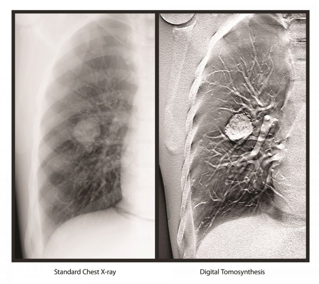
If you’ve ever been to the dentist, you’ve probably had X-rays taken. These X-rays produce pictures of your teeth that the dentist uses to help determine what type of dental care you need. Now imagine that instead of producing two-dimensional pictures of your teeth, a machine could actually help create three-dimensional models. Your dentist could look at each tooth from every angle — even take a cross-section of a tooth to look inside the structure. This technology is now possible thanks to something called digital tomography.Digital Tomography 101
Digital tomography (also known as computed tomography) combines classic X-ray technology with a computer system that processes the information. X-ray technology is used to take hundreds of images of your head. Computer programs can then read, interpret and even build virtual models based on these images.
Medicine has actually relied on this type of technology for years, in the form of a CT scanner. Cone-beam computed tomography (CBCT) is the dental equivalent of a conventional medical CT scan, with a few notable differences. X-ray radiation exposure with CBCT is significantly less than that for medical CT scans — up to 10 times less! The CBCT scans themselves take much less time to perform. And while medical CT scanners are quite large and usually restricted to hospitals or dedicated diagnostic imaging centers, CBCT scanners are smaller and are often available right in your dentist’s office.
Having a CBCT scan doesn’t hurt. Although there are several different models, most units are square-like machines with a chair. You will be asked to sit in a normal, upright-seated position with your chin resting in a chin cup while a C-shaped arm rotates around your head. While the images are being taken, you will need to keep your eyes closed and remain as still as possible. The entire process takes less than a minute.A Welcome Addition
Traditional X-rays have their place. But digital tomography takes dental imaging to the next level. It produces less distorted, sharper images. Using CBCT, your dentist can now distinguish between bone, tooth, nerves and soft tissue. CBCT scans can also identify possible tumors and other diseases that don’t appear on traditional X-rays. These capabilities translate into more informed dental treatment plans and more successful dental procedures.
One of the most common dental treatments relying on digital tomography is dental implants. Before placing a dental implant, your dentist can use the CBCT images to evaluate the quality and thickness of your jaw bone. Digital tomography also allows your dentist to locate areas that should be avoided when placing your dental implant.
In addition to helping in the placement of dental implants, computed tomography provides your dentist with a wealth of information which can be used to:
- Plan orthodontic treatment
- Evaluate temporomandibular joint disorders (TMJ)
- Diagnose jaw tumors
- Determine the presence of abnormal growths
- Diagnose periodontal disease
- Evaluate patients who have experienced dental trauma
The Full Story
Computed tomography does expose patients to radiation, as do all forms of dental X-rays. The level of exposure is equal to about 8 to 10 days’ worth of natural radiation exposure. That may sound like a lot, but compare it to the level of exposure from a medical CT scan, which exposes patients to about 10 months’ worth of naturally occurring radiation.
Since digital tomography is still a relatively new technology, it isn’t available everywhere. Plus, dentists must undergo special training to ensure correct interpretation of the images.

